
ポイント3心電図同期SPECT(QGS)画像の見方
1. 一般的なQGS画像表示上の、それぞれの画像の説明を図5に示す。
- ・拡張末期血流マップは、集積が低く描出されているが、これは壁厚が薄くなるために部分容積効果の影響でカウントが低下するためである。
- ・通常、血流評価は非同期画像で評価することを勧める。
- ・Motion(mm): 固定座標上の位置のずれ(距離)を表示。
- ・Thickening(%): 左室壁厚の変化率を表示。
- ・MotionとThickeningは基本的には一致する。ただし、乖離する症例もある。

図5 心電図同期SPECT(QGS)画面
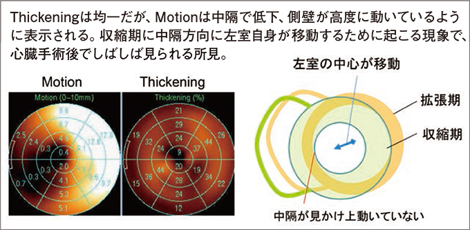
図6 心臓手術後(冠動脈バイパス)のThickeningとMotionマップ
2. 左室サイズ・LVEFと左室容積曲線
*表1 の正常値は小心臓例(QGS-EDV<50mL)を含んでいない。
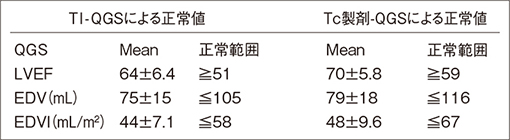
表1 東京女子医科大学における正常値
*なお、当院で左室造影(LVG)と比較検討したところ、LVEFは同等の数値であるが、QGSのEDV値はLVGに比べ低い値となっている。
EDV値は、後述する一過性虚血性内腔拡大(TID)の評価で指標として用いる。